
3D Digital Microscope vs. SEM: Which is more useful in your lab?

Published by Hirox USA | Digital Microscope Insights

If you work in materials science, electronics manufacturing, failure analysis, or advanced quality control, you have certainly had this conversation: should we be using a scanning electron microscope or a digital microscope for this?

 It is a fair question, and the honest answer is that both tools are genuinely powerful. But they solve different problems, operate under vastly different constraints, and carry quite different costs in time, money, and operational overhead. Choosing the wrong one for a given application does not just waste the budget. It slows down your team, degrades your data quality, and in the worst cases, lets critical defects slip through undetected.

 This post breaks down the real-world differences between SEM and 3D digital microscope so you can match the right instrument to the right job.

A Quick Primer: How Each Technology Works

scanning electron microscope works by firing a focused beam of electrons at a sample surface in a vacuum chamber. The way those electrons interact with the surface generates signals converted into high-resolution grayscale images. SEMs can achieve extraordinary magnification and reveal nanoscale surface detail invisible to optical systems.

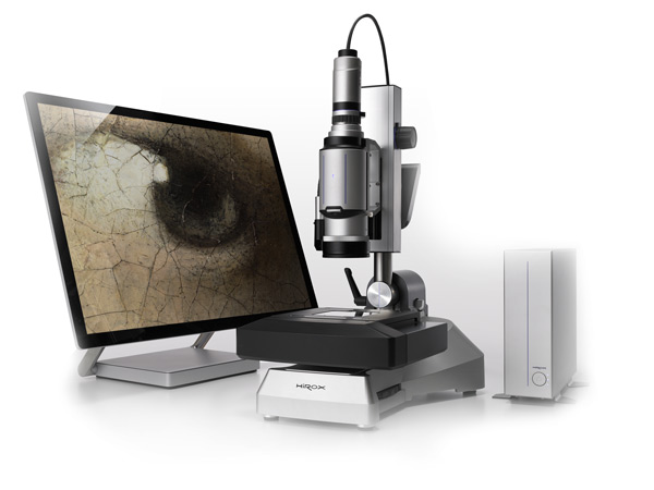
 3D digital microscope works with visible light. A high-resolution optical sensor captures reflected light across multiple focal planes, and the system constructs a true three-dimensional surface model — a full-color, measurable 3D dataset with actual topographical data at every point.

 Two fundamentally different physical approaches. Two fundamentally different sets of tradeoffs.

Sample Preparation: The Operational Cost Nobody Talks About Enough

SEM requires extensive sample preparation. Non-conductive materials must be sputter-coated with a thin layer of conductive metal before imaging. Samples must be sized to fit the vacuum chamber, which often means cutting, sectioning, or mounting larger parts. The entire imaging process takes place under high vacuum, ruling out wet samples or anything that degrades in that environment.

 For a research lab running dozens of similar samples per week, this workflow becomes routine. For a production QC environment where engineers need answers in minutes, not hours, it is a significant bottleneck.

 3D digital microscope requires no preparation for most samples. Parts are placed on the stage and imaged in air, at atmospheric pressure, exactly as they are. No coating. No sectioning. No vacuum pump down. A machined metal part comes off the production line and is under the lens within seconds — then goes back into the process undamaged and unchanged.

 For non-destructive testing requirements, standard in aerospace, medical device, and precision manufacturing, this is often a regulatory necessity, not just a convenience.

Magnification and Resolution: Understanding the Range

SEM’s headline advantage is magnification. At the high end, modern SEMs resolve features down to single-digit nanometers — a capability 3D digital microscope cannot match with visible light optics.

 But magnification range tells only part of the story. The more useful question is: what magnification range does your actual work require?

 For most industrial inspection, failure analysis, and quality control applications, the relevant range is 10x to 5,000x. Within that window, high-quality 3D digital microscopes perform extremely well — delivering sharp, full-color, three-dimensional images with sub-micron Z-axis resolution via confocal profilometry. They also offer seamless zooming without aperture changes, vacuum adjustments, or sample repositioning.

 At the extreme end — sub-100nm features, atomic-scale grain analysis — SEM remains the obvious choice. But for most industrial inspection tasks, 3D digital microscope covers the entire working range with significantly less friction.

Color Information: More Than Aesthetics

SEM images are grayscale. There is no color information in the electron interaction signals — it is an inherent physical limitation.

 For many applications, this matters significantly. Corrosion analysis, coating evaluation, and contamination identification all benefit from color data. A rust stain, an oil film, or a coating delamination are instantly recognizable in a full-color optical image, and require additional analytical steps like energy-dispersive X-ray spectroscopy to characterize in an SEM.

 3D digital microscopes capture full color natively. The surface looks like what you would see with your own eyes, just at much higher magnification with precise measurement data overlaid. For documentation and communicating findings to non-specialist stakeholders, this matters more than it might initially seem.

3D Measurement: The Data Dimension

A standard SEM produces a 2D image. Measurements on that image use projected dimensions — not the actual 3D geometry of the surface. Stereo SEM techniques can approximate 3D data, but the process is complex, slow, and far less accurate than dedicated profilometry.

 A 3D digital microscope generates a full surface height map as a baseline output. Every inspection automatically produces measurable topographical data: step heights, roughness parameters, volume measurements, cross-sectional profiles, and flatness values — all calculated from the 3D dataset and exportable in standard formats.

 For industries where surface finish specifications are part of the product definition — precision machined components, semiconductor wafers, medical implants, optical surfaces — this is the entire point of the inspection.

Throughput and Workflow Integration

When a production batch is on hold pending inspection results, every hour of instrument time translates directly into delay.

 3D digital microscopes are designed around production-floor realities. Motorized XY scanning stages capable of covering up to 1,500mm allow operators to create large-area panoramic scans automatically. Automated measurement routines can run unattended. Results are available in real time with no sample modification required.

 The difference in cycle time between the two technologies for a typical industrial inspection is not marginal. It can be measured in hours versus minutes.

Cost of Ownership: The Full Picture

Entry-level research SEMs typically start in the $80,000–$150,000 range, and high-performance field emission SEMs routinely exceed $500,000. Beyond acquisition cost, SEMs require dedicated electrical and HVAC infrastructure, vibration isolation, vacuum system maintenance, and in most cases a trained specialist operator.

3D digital microscopes require significantly less infrastructure and are designed to be operated by engineers, technicians, and quality personnel without specialized training.

For organizations weighing a first microscope investment, or looking to expand inspection capacity without scaling headcount, the total cost of ownership comparison often resolves the decision.

When to Choose SEM

Choose SEM when you need to characterize features below 100nm. Choose it when you need elemental composition data via EDS. Choose it when grain structure, crystallography, or nanoscale surface morphology is central to your analysis. For fundamental materials research, semiconductor process development, and nanoscience, SEM remains an essential instrument.

When to Choose 3D Digital Microscope

Choose 3D digital microscope when your work involves production inspection, non-destructive testing, or high-throughput quality control. Choose it when you need measurable surface data — roughness, step height, volume — not just images. Choose it when color information matters. Choose it when samples are large, geometrically complex, or need to be returned to service after inspection.

 Choose it when your team needs answers in minutes rather than hours, and when the people running the instrument are engineers and QC professionals rather than dedicated microscope specialists.

The Practical Answer: Both Have a Place

SEM and 3D digital microscopes are not really competing for the same applications. The organizations that use both typically have a clear division of labor — 3D digital microscope for routine inspection, documentation, and measurement, and SEM for edge cases that demand nanoscale resolution or compositional analysis.

 If your lab currently relies on SEM for work that does not require sub-100nm resolution or elemental analysis, there is a reasonable chance that a 3D digital microscope would serve most of those applications better — and free your SEM time for the cases where it is genuinely irreplaceable.


Curious what 3D digital microscope looks like applied to your specific inspection challenge? Contact the Hirox USA team to discuss your application or schedule a live demonstration.

 Hirox USA Inc. | 3D Digital Microscopes | NPS Confocal Systems | Inspection Services | www.hirox-usa.com


Why 3D Digital Microscopes Are Reshaping Quality Inspections Across Industries

Published by Hirox USA | Digital Microscope Insights

For decades, quality control teams, researchers, and engineers relied on traditional optical microscopes to inspect surfaces, measure features, and document findings. The process was slow, subjective, and limited — two-dimensional images, narrow depth of field, and tedious manual repositioning were simply the cost of doing business at the microscale.

Today, that paradigm is shifting fast. 3D digital microscopes has moved from a niche research tool to a mission-critical instrument across manufacturing, electronics, aerospace, forensics, conservation, and beyond. And if you want to understand why, the answer starts with what these systems can do that traditional microscopes simply cannot.

Seeing the Full Picture: What 3D Imaging Actually Means

When engineers talk about “3D microscope,” they’re not talking about a gimmick. They mean the ability to capture genuine topographical data — real height measurements across a surface — rather than a flat, two-dimensional photograph of it.

This distinction matters enormously in practice. Consider a weld bead on a critical aerospace component. A 2D image can show cracks, discoloration, or porosity — but it tells you nothing about depth, volume, or surface roughness. A 3D digital microscope captures all of that simultaneously, generating measurable data that can be documented, compared across batches, and shared with a team thousands of miles away.

The result isn’t just a better image. It’s a complete inspection record.

The Rotary-Head Advantage: 360° Around Complex Parts

One of the most persistent challenges in microscopy has always been sample geometry. Flat surfaces are easy. But what about threaded fasteners, connector pins, turbine blades, or surgical instruments? Traditional microscopes are essentially designed for flat samples — anything with relief, curvature, or undercuts becomes a frustrating puzzle of repositioning and compromise.

Hirox’s patented 360° rotary-head technology solves this directly. The motorized rotation system allows the lens to orbit completely around a sample, capturing every angle without ever touching or disturbing the part. The result is what Hirox calls a “helicopter view” — a full circumferential record of a component’s surface that would be impossible to achieve with a conventional setup.

For industries where connector integrity, thread quality, or edge geometry are safety-critical, this capability isn’t a luxury. It’s a requirement.

Speed Without Sacrifice: Why Throughput Matters

Laboratory throughput is often the unsung hero of quality programs. A microscope that produces beautiful images but requires twenty minutes of setup per sample is a bottleneck that erodes the value of every inspection it performs.

Modern 3D digital microscopes are engineered around the reality that inspection teams have real production schedules to meet. Wide-range zoom lenses, motorized XY scanning stages spanning up to 1,500mm, and automated stitching capabilities allow operators to cover large sample areas quickly — without sacrificing the resolution detail that makes the inspection meaningful.

When a single system can move from a macro survey of a circuit board to a sub-micron surface analysis of a solder joint in seconds, the conversation around inspection capacity changes completely.


Confocal Profilometry: When Nanometers Count

For applications where surface roughness is measured in nanometers rather than microns — semiconductor fabrication, precision optics, advanced coatings — standard digital microscopy isn’t enough. This is where confocal chromatic profilometry enters the picture.

The Nano Point Scanner (NPS) approach uses white light confocal technology to achieve submicron Z-axis precision, capturing surface profiles with extraordinary accuracy. The measurement results are ISO-certified and can be automatically compiled into standardized reports — critical for organizations operating under strict quality management systems or regulatory frameworks.

In an era where component tolerances are shrinking and failure analysis demands are growing, the ability to measure with this level of confidence is a genuine competitive differentiator.


Documentation That Works as Hard as the Instrument

The best inspection data in the world is useless if it lives only in an operator’s memory or a folder of unlabeled images. Modern quality programs demand traceability — the ability to document when a part was inspected, by whom, under what conditions, and with what result.

A 3D digital microscope supports this natively. Automated image analysis, integrated measurement reporting, and structured data export mean that every inspection generates a defensible, reviewable record. For industries operating under FDA oversight, ISO certification, or aerospace quality standards, this capability alone can justify the investment.

Applications Spanning Every Industry

The versatility of a 3D digital microscope is one of its most undera ppreciated qualities. The same platform that inspects microelectronics solder joints on Monday can be used for:

  • Failure analysis of fractured metal components
  • Surface characterization of medical implants and devices
  • Authentication and documentation of fine art and cultural artifacts
  • Weld and coating inspection in automotive and heavy manufacturing
  • Forensic analysis of toolmarks, fibers, and trace evidence
  • R&D surface characterization in materials science and polymer research

This breadth is possible because the core capability — high-resolution, quantitative, non-contact surface imaging — is fundamentally useful anywhere that surfaces matter and precision is required.

The Digital Microscope Difference: A Shift in How Teams Work

Perhaps the most transformative aspect of a modern digital microscope isn’t any single technical capability — it’s the way these systems change how inspection teams collaborate and communicate.

When a microscope produces a 3D dataset rather than a photograph, that data can be shared, annotated, and reviewed by colleagues in different departments or different continents. Inspectors can hand off a finding not as a subjective description but as a measurable, three-dimensional record. Engineers reviewing a failure don’t have to travel to a lab — they can examine the surface data in detail from their own workstations.

This shift toward data-driven, collaborative inspection workflows is one reason 3D digital microscopes has moved so quickly from specialized research tool to mainstream quality instrument.


Hirox: Four Decades of Innovation at the Microscale

Hirox invented the first video microscope more than 40 years ago — a milestone that established the foundational concept of digital imaging at the microscale. Since then, the company has continued to advance the field through innovations like the 360° rotary-head system, motorized scanning stages, and the NPS confocal profilometry platform.

Today, Hirox USA supports customers across industries with not just best-in-class instruments, but a full suite of services including calibration and maintenance, advanced operator training, inspection and measurement consulting, and application-specific video documentation. The goal has always been the same: give technical teams the tools and knowledge to see more, measure more, and decide with confidence.

Ready to see what a 3D digital microscope can reveal in your application? Contact Hirox USA to schedule a demonstration or speak with an applications specialist.

Hirox USA Inc. | 3D Digital Microscopes | NPS Confocal Systems | Inspection Services www.hirox-usa.com