Worried that switching to a new digital microscope means downtime and retraining? Here’s why upgrading to a Hirox system is faster than you’d expect.
Ask any lab manager or QC lead why they’re still working with an aging inspection setup, and the answer is rarely about the equipment itself. It’s about the switch. New instruments mean downtime, retraining, revalidated workflows, and the nagging fear that the “upgrade” will slow the team down before it speeds anything up.
That concern is understandable — and, in the case of a Hirox digital microscope, largely unfounded. Hirox systems were engineered from the start to lower the barrier between “we should modernize” and “we’re up and running.” Here’s what the transition actually looks like.
No Eyepiece, No Learning Cliff
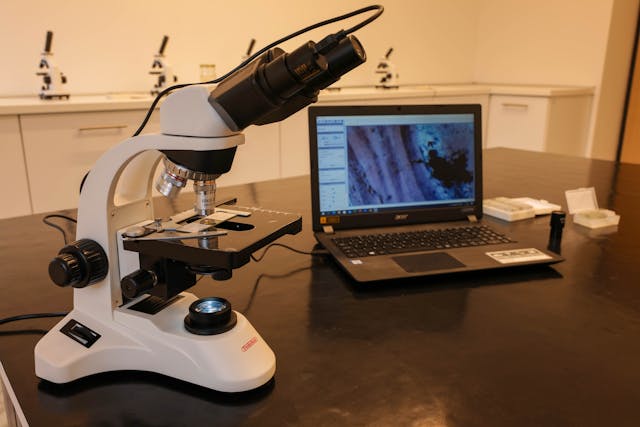
The first thing operators notice about a digital microscope is what’s missing: the eyepiece. Instead of hunching over binoculars, your team views a live, high-resolution image on a large monitor — a far more natural way to work, and one that makes training dramatically faster.
With the Hirox HRX-02, a 5-megapixel CMOS sensor delivers crystal-clear images at 50 frames per second on a standard 4K display. Everyone in the room sees the same image at the same time, which means a senior inspector can walk a new hire through a sample in minutes rather than trading turns at an eyepiece. Features like real-time multi-focus — which instantly builds a fully focused image even on samples with significant height variation — remove the depth-of-field frustration that makes traditional microscopes feel like a specialist’s craft. Anyone on the team can produce a usable, fully focused image on day one.
It Runs on Hardware You Already Have
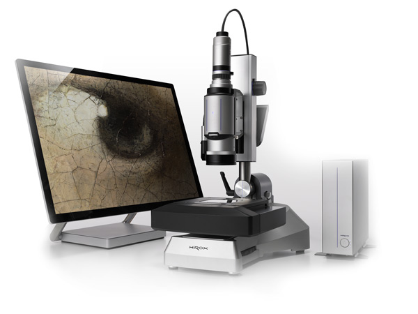
Legacy inspection systems often arrive as sealed, all-in-one towers: proprietary computer, proprietary monitor, proprietary headaches. The current generation of Hirox systems takes the opposite approach. The HRX platform connects to a standard Windows PC over USB and takes full advantage of the 4K displays most facilities already own.
That has practical consequences for an upgrade. IT doesn’t need to certify exotic hardware. The microscope fits into your existing network, storage, and documentation workflows. And when you eventually refresh your PCs, your microscope simply comes along for the ride instead of becoming stranded on obsolete hardware.
Smart Hardware That Prevents Mistakes
One of the quiet anxieties of any equipment transition is operator error during the adjustment period. Hirox has engineered much of that risk out of the system. Intelligent magnetic lens adapters with embedded RFID chips let the software instantly recognize which lens is attached — no manual calibration entry, no mismatched settings, no measurements taken at the wrong magnification profile.
On the HRX-02, built-in intelligence goes a step further, automatically selecting optimal observation settings based on the sample and scene. An ergonomic joystick puts lighting, stage movement, zoom, and image capture under one hand. The result is a system where the “expert knowledge” lives in the instrument, not in a binder of tribal knowledge that walks out the door when a senior technician retires.
A Modular System That Grows With You
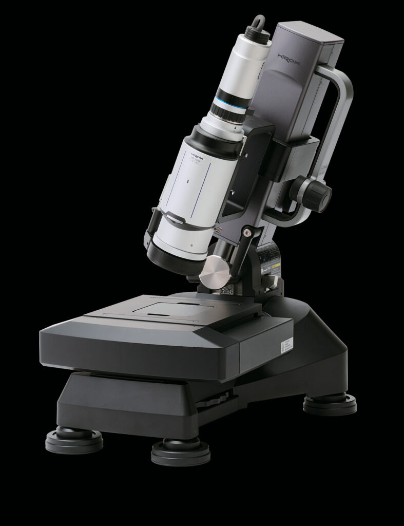
Upgrading to Hirox isn’t an all-or-nothing leap. The platform is deliberately modular: a single system spans magnifications from macro observation up to 10,000x simply by changing lenses. Motorized XY stages range from 50 × 50 mm up to 1,000 × 1,000 mm for automated scanning and large-area 3D stitching. The patented 360° rotary-head adapter adds full circumferential inspection of threaded, curved, or complex parts — without ever touching the sample.
And when your measurement requirements advance, the system advances with you. The NPS white-light confocal add-on brings sub-micron surface profilometry onto the same platform. In other words, the digital microscope you install this quarter is the foundation for capabilities you may not need until next year — no forklift replacement required
Support That Starts Before You Buy
Hirox USA treats the upgrade as a process, not a shipment. It begins with a demonstration — either a live web demo or an on-site demo at your facility, using your actual samples. That means you know exactly how the system performs on your parts before a purchase order is ever written.
After installation, the support continues: operator training, calibration and maintenance services, sample analysis, and application consulting are all part of the Hirox USA offering. Teams aren’t handed a manual and wished luck; they’re brought up to speed by application specialists who work with these systems every day.
Four Decades of Making Microscopes Easier
There’s a reason the transition feels smooth: it’s been refined for a long time. Hirox invented the first video microscope more than 40 years ago and has been designing lenses since 1920. Every generation of the platform — through today’s HRX-01 and HRX-02 — has been shaped by the same philosophy: superior optics first, with usability designed in rather than bolted on.
That heritage matters when you’re the one signing off on an equipment change. You’re not betting your inspection workflow on an unproven design. You’re stepping onto a platform that thousands of labs, factories, forensic units, and conservation studios have already adopted — most of them faster than they expected.
Ready to See It on Your Samples?
The easiest way to evaluate an upgrade is to watch a Hirox digital microscope work on the exact parts your team inspects every day. Contact Hirox USA to schedule a web demo or an on-site demonstration at your facility.