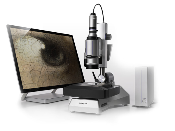
Published by Hirox USA | Digital Microscope Insights
For decades, quality control teams, researchers, and engineers relied on traditional optical microscopes to inspect surfaces, measure features, and document findings. The process was slow, subjective, and limited — two-dimensional images, narrow depth of field, and tedious manual repositioning were simply the cost of doing business at the microscale.
Today, that paradigm is shifting fast. 3D digital microscopes has moved from a niche research tool to a mission-critical instrument across manufacturing, electronics, aerospace, forensics, conservation, and beyond. And if you want to understand why, the answer starts with what these systems can do that traditional microscopes simply cannot.
Seeing the Full Picture: What 3D Imaging Actually Means
When engineers talk about “3D microscope,” they’re not talking about a gimmick. They mean the ability to capture genuine topographical data — real height measurements across a surface — rather than a flat, two-dimensional photograph of it.
This distinction matters enormously in practice. Consider a weld bead on a critical aerospace component. A 2D image can show cracks, discoloration, or porosity — but it tells you nothing about depth, volume, or surface roughness. A 3D digital microscope captures all of that simultaneously, generating measurable data that can be documented, compared across batches, and shared with a team thousands of miles away.
The result isn’t just a better image. It’s a complete inspection record.
The Rotary-Head Advantage: 360° Around Complex Parts
One of the most persistent challenges in microscopy has always been sample geometry. Flat surfaces are easy. But what about threaded fasteners, connector pins, turbine blades, or surgical instruments? Traditional microscopes are essentially designed for flat samples — anything with relief, curvature, or undercuts becomes a frustrating puzzle of repositioning and compromise.
Hirox’s patented 360° rotary-head technology solves this directly. The motorized rotation system allows the lens to orbit completely around a sample, capturing every angle without ever touching or disturbing the part. The result is what Hirox calls a “helicopter view” — a full circumferential record of a component’s surface that would be impossible to achieve with a conventional setup.
For industries where connector integrity, thread quality, or edge geometry are safety-critical, this capability isn’t a luxury. It’s a requirement.
Speed Without Sacrifice: Why Throughput Matters
Laboratory throughput is often the unsung hero of quality programs. A microscope that produces beautiful images but requires twenty minutes of setup per sample is a bottleneck that erodes the value of every inspection it performs.
Modern 3D digital microscopes are engineered around the reality that inspection teams have real production schedules to meet. Wide-range zoom lenses, motorized XY scanning stages spanning up to 1,500mm, and automated stitching capabilities allow operators to cover large sample areas quickly — without sacrificing the resolution detail that makes the inspection meaningful.
When a single system can move from a macro survey of a circuit board to a sub-micron surface analysis of a solder joint in seconds, the conversation around inspection capacity changes completely.
Confocal Profilometry: When Nanometers Count
For applications where surface roughness is measured in nanometers rather than microns — semiconductor fabrication, precision optics, advanced coatings — standard digital microscopy isn’t enough. This is where confocal chromatic profilometry enters the picture.
The Nano Point Scanner (NPS) approach uses white light confocal technology to achieve submicron Z-axis precision, capturing surface profiles with extraordinary accuracy. The measurement results are ISO-certified and can be automatically compiled into standardized reports — critical for organizations operating under strict quality management systems or regulatory frameworks.
In an era where component tolerances are shrinking and failure analysis demands are growing, the ability to measure with this level of confidence is a genuine competitive differentiator.
Documentation That Works as Hard as the Instrument
The best inspection data in the world is useless if it lives only in an operator’s memory or a folder of unlabeled images. Modern quality programs demand traceability — the ability to document when a part was inspected, by whom, under what conditions, and with what result.
A 3D digital microscope supports this natively. Automated image analysis, integrated measurement reporting, and structured data export mean that every inspection generates a defensible, reviewable record. For industries operating under FDA oversight, ISO certification, or aerospace quality standards, this capability alone can justify the investment.
Applications Spanning Every Industry
The versatility of a 3D digital microscope is one of its most undera ppreciated qualities. The same platform that inspects microelectronics solder joints on Monday can be used for:
- Failure analysis of fractured metal components
- Surface characterization of medical implants and devices
- Authentication and documentation of fine art and cultural artifacts
- Weld and coating inspection in automotive and heavy manufacturing
- Forensic analysis of toolmarks, fibers, and trace evidence
- R&D surface characterization in materials science and polymer research
This breadth is possible because the core capability — high-resolution, quantitative, non-contact surface imaging — is fundamentally useful anywhere that surfaces matter and precision is required.
The Digital Microscope Difference: A Shift in How Teams Work
Perhaps the most transformative aspect of a modern digital microscope isn’t any single technical capability — it’s the way these systems change how inspection teams collaborate and communicate.
When a microscope produces a 3D dataset rather than a photograph, that data can be shared, annotated, and reviewed by colleagues in different departments or different continents. Inspectors can hand off a finding not as a subjective description but as a measurable, three-dimensional record. Engineers reviewing a failure don’t have to travel to a lab — they can examine the surface data in detail from their own workstations.
This shift toward data-driven, collaborative inspection workflows is one reason 3D digital microscopes has moved so quickly from specialized research tool to mainstream quality instrument.
Hirox: Four Decades of Innovation at the Microscale
Hirox invented the first video microscope more than 40 years ago — a milestone that established the foundational concept of digital imaging at the microscale. Since then, the company has continued to advance the field through innovations like the 360° rotary-head system, motorized scanning stages, and the NPS confocal profilometry platform.
Today, Hirox USA supports customers across industries with not just best-in-class instruments, but a full suite of services including calibration and maintenance, advanced operator training, inspection and measurement consulting, and application-specific video documentation. The goal has always been the same: give technical teams the tools and knowledge to see more, measure more, and decide with confidence.
Ready to see what a 3D digital microscope can reveal in your application? Contact Hirox USA to schedule a demonstration or speak with an applications specialist.
Hirox USA Inc. | 3D Digital Microscopes | NPS Confocal Systems | Inspection Services www.hirox-usa.com